Watching the brain adapt to light and dark

Written by: Ken Estrellas

Original Article: Wang et al. PLoS Biology 2020
The Gist of It
Throughout the world, animals of all shapes and sizes demonstrate behaviors linked to cycles of light and dark, from sleeping and wakefulness in humans to navigation in butterflies. These patterns are described as circadian rhythms, processes that are governed by external factors such as light and temperature and tend to cycle every 24 hours. Scientists have studied circadian rhythms in a variety of ways, but the researchers in this particular study have developed what might be the most sophisticated method yet – watching individual cells in a living animal turning genes on and off in cycles of light and dark. Wang and colleagues used zebrafish that were genetically modified to emit a fluorescent protein, Venus-NLS-PEST (VNP), in cells that also have the gene nr1d1, a circadian clock gene, turned on. By putting the zebrafish in a special circulating water bath and using a particular microscope known as a “two-photon microscope,” the authors were able to track and film cells that had increasing levels of VNP in the dark and decreasing levels in the light, which meant that the cells had the circadian clock gene turned on and then off. Most of these cells were located in the pineal gland, a part of the brain in most vertebrates that produces melatonin, a hormone which controls sleep patterns. Specifically, these cells were identified as melatonin-secreting cells that were light sensitive – similar to the ones in our eyes! Finally, in order to demonstrate that light/dark cycles were necessary for these cells to develop, a different set of fish were raised under constantly dark conditions. Without the light/dark cycle, the fish showed decreased levels of fluorescence in regions of the brain that would light up in fish raising with normal light/dark cycles. Overall, the authors of this study appear to have successfully created a unique method for studying circadian rhythm and its effects on living creatures.
Screen Shot 2020-03-29 at 5.11.02 PM

With this new system, scientists can watch how certain brain cells react to light and dark – inside of a living animal!

The Nitty Gritty
In this study, Wang et al. report the development of a novel zebrafish-based model to study circadian rhythm. Transgenic zebrafish lines were generated with expression of Venus-NLS-PEST (VNP) linked to both the proximal and distal promoter regions of nr1d1, a key circadian clock gene which expresses a transcription factor linked to the circadian BMAL1/CLOCK axis. Quantitative PCR revealed oscillating expression of both VNP and nr1d1 roughly linked to 12-hour day/night cycles between 3.5-7.5 days post-fertilization (dpf), with highest levels of expression observed just before “dawn” and lowest levels just before “dusk.” This quantitative assessment of expression was visualized by mounting anesthetized live zebrafish in low melting point agarose and a circulating water bath, then monitoring VNP fluorescence using two-photon microscopy. The most prominent reporter expression as measured by fluorescence intensity was observed in the pineal gland, with subsequent development observed in the optical tectum at 5.5 dpf and cerebellum at 6.5 dpf. When nr1d1:VNP zebrafish were crossed with a second line linking expression of aanat2 with mRFP, significant co-localization was observed, indicating that the cells lighting up with nr1d1:VNP were melatonin-synthesizing photoreceptor cells. Further crosses and co-localization analysis with xops:nfsB-mCherry and lws2:nfsB-mCherry lines revealed that these cells also co-express markers for rod-like and cone-like photoreceptor cells, respectively. Rod-like cells were shown to have higher levels of baseline nr1d1:VNP expression than non-rod like cells were, but single-cell analysis revealed that both cell types maintain oscillations linked to light-dark cycles. Finally, raising a subset of fish in continuously dark conditions from 0-3.5 dpf significantly dampened nr1d1:VNP oscillation amplitudes, indicating the necessity of light-dark cycling on the successful development of circadian expression. Conversely, normally-raised fish subsequently transferred into dark conditions maintained their oscillations. Taken together, these results indicate the successful generation of a novel reporter line to study circadian rhythms with live fluorescence imaging at a higher single-cell resolution compared to previously developed bioluminescence-based lines.
Original Research Article: Wang, H., et al. “Single-cell in vivo imaging of cellular circadian oscillators in zebrafish.” PLoS Biol 18.3 (2020): e3000435.

Overcoming barriers: How Zika virus gets to the brain

Written By: Rebecca Tweedell

The Gist of It:
In 2015 and 2016, the world was closely following the outbreak of a mosquito-borne virus called Zika. While these outbreaks have since faded from the forefront, with very few if any cases being detected currently, there are still a lot of unknowns about this virus. One thing we do know is that Zika virus can cause fetal microcephaly, or swelling of the brain in unborn babies. To do this, the virus must somehow be able to get to the brain. This is especially challenging because there are 2 key lines of defense working to keep viruses out, the placenta and the blood-brain barrier. The placenta is formed during pregnancy and allows oxygen and nutrients to be passed from the mother to the baby while blocking the passage of toxic materials and viruses. The blood-brain barrier exists in both babies and adults and serves as the boundary to keep the circulating blood (and any bad things that might be circulating with it) away from the brain. Since we know that the virus can get into the brain, we know these barriers must be failing, but how this happens is still a big question. Researchers at National Yang-Ming University in Taiwan recently took a major step toward solving this mystery. They studied how the virus passed through placental barrier cells and brain-derived barrier cells in the lab. They discovered that the virus actually crosses these barriers in different ways. Zika virus disrupted the ability of the placental cells to form strong junctions between themselves, weakening the physical structure of the barrier. This was not the case with the brain-derived cells, which maintained the physical structure of the barrier in spite of the virus. But the virus was still able to move through both types of barriers by going directly through the cells through a process known as transcytosis (which literally means being transported across the interior of the cell). Excitingly, blocking transcytosis with inhibitors was able to keep Zika virus from getting through the barriers. Understanding how the virus is able to get through these barriers is important for being able to stop it, and this study helped us take a big step in the right direction.
iceberg

Zika virus can pass through placental barriers by going between cells, breaking their tight junctions, and the virus can pass through both placental and brain barriers by using transcytosis to go directly through the cells to get to the brain.

The Nitty Gritty:
Chiu et al. used the human placenta trophoblast cells JEG-3 and human brain-derived endothelial cells hCMEC/D3 to create an in vitro model to study interactions with Zika virus. They found that Zika virus could infect both of these cell types when the cells were grown in a monolayer. Using a transwell barrier assay, with a monolayer of JEG-3 or hCMEC/D3 cells in the insert and Vero cells in the bottom chamber, the researchers found that Zika virus was able to pass through the barrier formed by the cells, moving from the portion of the well above the JEG-3 or hCMEC/D3 cells to infect the Vero cells below. When passing through the JEG-3 placental cells, Zika virus damaged the integrity of the membrane, allowing FITC-dextran to pass through. However, this did not occur with the hCMEC/D3 cells. This was likely due to tight junction disruption; the researchers found that the expression of ZO-1 and occludin was decreased in JEG-3 cells following exposure to Zika virus, while this did not occur in hCMEC/D3 cells. After treatment with a proteasome inhibitor MG132, the expression of ZO-1 and occludin was rescued, suggesting that the disruption of tight junctions occurs through the proteasomal degradation pathway. Finally, the authors sought to understand whether Zika virus can use transcytosis to traverse the placental and brain-derived barrier cells. Using a fluorescently labeled virus, they measured the fluorescence in the basal chamber of a transwell following culture of the virus with the monolayer barrier in the insert at 4°C and 37°C, as transcytosis is inhibited at 4°C. They found that the amount of virus that traversed the barrier at 4°C was significantly reduced compared to the amount that traversed at 37°C, while there was no difference in the amount of FITC-dextran that could traverse. Based on the finding that the virus is able to traverse the cells by transcytosis, the authors tried to block the virus using inhibitors of endocytosis and intracellular trafficking (Nystatin, chlorpromazine, dimethyl amiloride, and colchicine). Each inhibitor reduced the amount of virus that made it through the monolayer barrier. Overall, Chiu et al. found that Zika virus crosses placental and brain-derived barrier cells in culture by transcytosis and that the virus disrupts the integrity of tight junctions to weaken the placental cell barrier in the transwell system. These findings pave the way for discovering ways to inhibit viral traversal and prevent fetal infection and encephalitis.
Original Research Article: Chiu, C.-F., et al. “The mechanism of the Zika virus crossing the placental barrier and the blood-brain barrier.” Front Microbiol (2020).

Does a tense environment affect behavior? A synthetic extracellular matrix may help answer that question

Written By: Kaitlyn Sadtler

Original Article: Davidson et al. Acta Biomaterialia 2020
The Gist of It:
Just like we take in information from our environment and change our behavior (like putting a coat on when it’s cold outside), cells take in information from their environment – the extracellular matrix. The cues they receive can cause them to change their environment by secreting different proteins, making the matrix stiffer or softer (just like we can turn on the A/C or put another log on the fire to make it cooler or warmer). Stiffening and densifying of the extracellular matrix is called fibrosis, and this process contributes to numerous disorders, including liver cirrhosis, cardiac dysfunction, skin scarring, and lung fibrosis. Understanding how our cells sense changes in the stiffness of the matrix around them and how that affects disease progression is necessary for the development of new therapeutics. Researchers from the University of Michigan Ann Arbor have developed a new material and platform to study this phenomenon in the laboratory. Davidson and colleagues synthesized a fibrous biologic matrix out of dextran vinyl sulfone (DexVS). The researchers were able to spin these fibers onto a plate to create a sort of cellular hammock, where the cells would feel just the stiffness of the material and not the plastic bottom of a petri dish. With these fibers, they were able to look in a highly controllable manner at how cells interacted with stiffer and softer matrices. In contrast to what had been seen when the interaction between cells and the matrix was studied using other methods, like looking at solid synthetic gels (jello-like), they found that stiffer materials made cells less fibrotic than softer materials did. Using these models, scientists will be able to probe interactions between cells and fiber-based matrices that are more similar to the natural extracellular matrix and compare to previous studies which used fully synthetic hydrogels that do not have fibers. Moving forward, these scientists have developed ways to modify these materials with different proteins and change the way that the cells interact with the matrix. Further research like this could expand our understanding of what happens to our cells in stiff environments, which could lead to therapeutics for fibrotic diseases.
iceberg

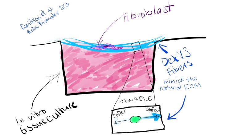
New Dextran Vinyl Sulfone (DexVS) fibers are tunable, which means scientists can alter the stiffness of each fiber, along with the stiffness of the overall synthetic extracellular matrix they have created. Using a draped matrix, cells in contact with the fibers will only feel stiffness of those fibers, and not the plastic around them.

The Nitty Gritty:
Previously, researchers had developed an electrospun methacrylated dextran (DexMA) to study matrix stiffness in vivo. However, DexMA fibers degraded in vitro due to hydrolytic cleavage of the ester bonds in the polymeric matrix within days of exposure to cell culture media. Here, the researchers presented a dextran vinyl sulfone (DexVS) that lacks the sensitive ester bonds and can still be functionalized via Michael-type addition and crosslinked with a photoinitiator (in this case, lithium phenyl-2,4,6-trimethylbenzoylphosphinate, LAP). They tested the stiffness of these materials by changing the concentrations of photoinitiator or duration of UV exposure. They further functionalized these materials with cyclic RGD (cRGD) for cell attachment or with methacrylated heparin to promote association of cell-secreted matrix with the synthetic polymer fibers. DexVS fibers were spun onto a multi-well plate to create a suspended surface with mechanical properties independent of the tissue culture plastic. Culturing normal human lung fibroblasts (NHLFs) on stiffer and softer matrices revealed that softer fibrous matrices, in the presence of pro-fibrotic soluble transforming growth factor beta-1 (TGFβ1), induced higher levels of myofibroblast-associated alpha smooth muscle actin (αSMA) than did stiffer matrices. This is contradictory to studies performed with solid hydrogels, such as poly(ethylene glycol) (PEG)-based hydrogels, that suggested stiffer matrices induce more of the fibrotic myofibroblast phenotype. Further research using multiple model systems in vitro and comparison to in vivo samples will hone in vitro modeling to pave the way for understanding mechanistic biology, modeling disease, and screening novel therapeutics.
Original Research Article: Davidson, C.D., et al. “Myofibroblast activation in synthetic fibrous matrices composed of dextran vinyl sulfone.” Acta Biomaterialia (2020).