
Written By: Ritu Raman
Original Article: Can et al. 2017.
The Gist of It:
When you think of an electronic circuit, you probably think wires, little metal pieces, and flashing LEDs. A new trend engineers have been exploring is building biological tissues that mimic the properties of electronic components. Designing circuits that are partly or completely biological is the goal of the emerging field of “biocomputing”, or using biological systems for information processing, and has many potential applications including smart prosthetics and implantable sensors. In this paper, Can et al created a biological system that mimicked the properties of an electronic diode. A diode is an electrical component that only allows current to flow in one direction. A mechanical analogy for this is a check valve in a piece of tubing that only allows water within the tubing to flow one way but not the other. Can et al created a biological diode using two types of cells patterned next to one another: cardiac muscle cells and cardiac fibroblasts (connective tissue). Cardiac muscle cells are “excitable”, which means they respond to external electrical signals, and can transmit that electrical signal to nearby cells through “gap junctions”, channels that physically connect adjacent cells. By contrast, cardiac fibroblasts are nonexcitable cells, so they can’t respond to external electrical signals. They can, however, receive electrical signals through gap junctions and propagate them to nearby cells. As a result, when the muscle cells are excited by an electrical signal, the electrical pulse travels through the muscle cells and through adjacent fibroblasts. However, when an electrical signal is delivered to the cardiac fibroblasts, the pulse cannot travel. This directional control over the pulse’s travel creates a “living” biological diode (Fig. 1). In time, I’m hoping this team and others develop other bio-electrical components, so we can start building biological circuits from scratch!
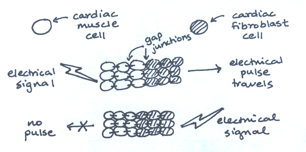
Building electrical circuits using living cells.
